Echocardiogram, Stress Echocardiogram, Echo Bubble Study
Echocardiogram
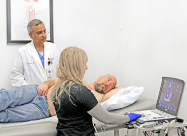
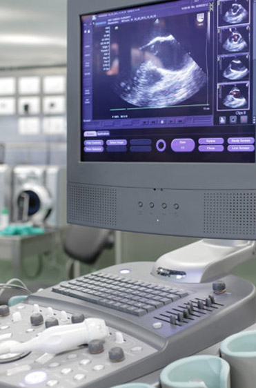
An echocardiogram uses high frequency sound waves to produce pictures of the heart. The ultrasound technician guides a small hand-held device called a transducer over the chest, that emits soundwaves. These sound waves bounce off internal substances and structures (in this case the heart), creating echoes that are picked up by the transducer. These are converted into moving images of the heart’s chambers, valves, blood vessels and pumping action. Combined with doppler technology, the speed and direction of the heart’s blood flow can also be measured at the same time.
Echocardiograms can help detect many aspects of heart disease including tumors, abnormal holes between the heart chambers (ASD & PFO), blood clots, and issues with the heart’s blood vessels, valves and pericardium (the heart’s outer lining).

Stress Echocardiogram
A stress echocardiogram, or stress echo test, assesses how well the heart is functioning when it is working harder due to physical stress. For comparison reasons, a resting echocardiogram is first administered. An ultrasound technician guides a small hand-held device called a traducer over the chest. It transmits soundwaves that are converted to moving images of the heart on the computer screen. Next, the patient walks on a treadmill while the heart rhythm and blood pressure are monitored. When the heart rate increases to a certain point, a second set of echocardiogram images are taken. This test shows how efficiently your heart pumps blood, can detect heart issues that may not show up at rest, and can measure the severity of heart issues such as a blockage. In cases where the patient is unable to walk on a treadmill, an IV medication can be given instead that will mimic the effects of exercise to increase the heart rate.

Echo Bubble Study
An echo bubble study can be performed during an echocardiogram. During the echocardiogram, a saline solution is rapidly mixed with air to produce tiny bubbles, and is then injected into the patient’s vein in their arm through an IV. The bubbles circulate up the vein into the right side of the heart, showing up on the echocardiogram. The bubbles should normally then be filtered out by the lungs. However, if these bubbles appear on the left side of the heart, this indicates that there is a hole (an ASD or PFO) between the heart’s two upper chambers (the left and right atria).


