Peripheral Artery Disease
Peripheral artery disease (PAD) is a disease in which a fatty substance called plaque builds up inside the peripheral arteries. Peripheral arteries bring oxygen-rich blood to your arms and legs—the extremities of the body. When plaque builds up in these arteries, it creates a narrowing or blockage, limiting the blood flow supply to your arms or legs. In its early stages, PAD causes painful cramping while you walk. If the disease progresses, it may cause variety of other symptoms that could include:
Your risk of PAD increases as you get older. Risk factors for PAD include:
Diagnosis
To diagnose PAD, we start with a thorough physical exam. We then will use a variety of tests to further diagnosis PAD which include:
Ankle-brachial index (ABI)
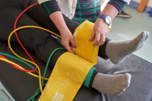
Ankle-brachial index (ABI) is a test for PAD, that compares the blood pressure in the leg to the blood pressure in the arm. If the blood pressure in the arm is higher than the blood pressure in your legs, this could be an indication of decreased blood flow in your legs known as Peripheral Artery disease or PAD.

Arterial Duplex Ultrasound / Arterial Doppler Ultrasound
An arterial duplex ultrasound, also known as an arterial doppler ultrasound, shows blood flow through the arteries. The ultrasound technician presses and moves a small hand-held device that produces soundwaves, over the area being examined. These sound waves create color images of the veins and arteries. This allows us to measure blood flow in the arteries, to determine if there is a blockage. Based on the findings of this test, your doctor will determine if further testing or treatment are necessary.
Peripheral Angiogram / Peripheral Vascular Catheterization
A peripheral angiogram, also known as a peripheral vascular catheterization, is a specialized test done under X-ray, that shows the blood flow through the peripheral arteries. The peripheral arteries are responsible for supplying oxygenated blood to your arms and legs. During the procedure, your doctor makes a small incision in the wrist (radial artery) or groin (femoral artery) and advances a long thing tube, called a catheter, through the artery leading up into your peripheral artery in your leg. Your doctor then injects a special fluid called contrast media, or contrast dye, through the catheter into your artery’s blood stream.
The contrast dye allows the x-ray to show real time moving images of the blood flow through the peripheral arteries. The doctor uses these images to identify the precise location of any blockages in the arteries and the extent of plaque buildup. Based on the findings of this test, your doctor is able to determine if further treatment such as a peripheral angioplasty or peripheral stenting is needed to fix the lack of blood supply.
Treatment
Although PAD cannot be cured, we can reduce symptoms and prevent future problems such as heart attack or stroke. We may start by recommending lifestyle changes such as exercising, weight loss, and smoking cessation. We may also prescribe medications. In certain cases, we offer a variety of minimally invasive and open surgical procedures that can help. Minimally invasive procedures include:
Peripheral Angioplasty / Peripheral Vascular Intervention (PVI)
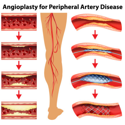
A peripheral angioplasty, also known as peripheral vascular intervention (PVI), is a minimally invasive procedure that can improve blood flow to the pelvis or legs when a person has peripheral artery disease. Our doctor advances a catheter (a long narrow tube) with a tiny balloon on its tip up into the artery until it reaches the site that the plaque buildup is causing the blockage. A wire is then passed through the blocked area that allows the doctor to use the equipment necessary to fix the blockage.
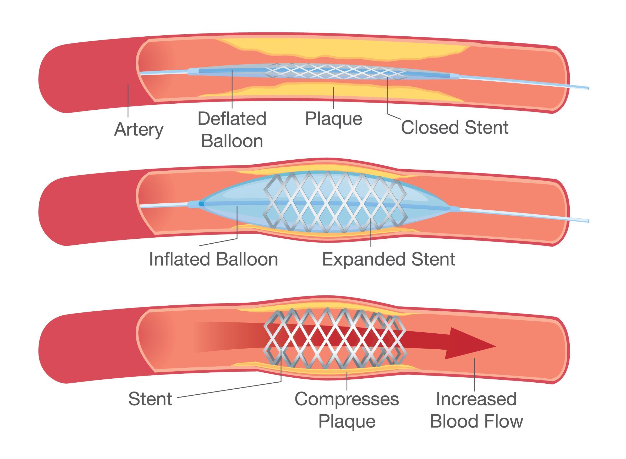
The equipment can include balloons, stents or other more specialized devices depending on the nature and severity of the disease. These devices are then passed along the wire to the desired location. The balloon is inflated to compress the plaque against the artery wall, opening up the passageway and restoring blood flow. The balloon is then deflated and removed. In most cases, your doctor may determine that a stent needs to be inserted where the blockage was to keep the artery open after the balloon is removed.

Stents
A stent is a tiny wire mesh scaffold that helps keep the artery open to maintain blood flow into the heart. In many cases, a stent is placed during an angioplasty. The doctor inserts a balloon catheter covered with the unexpanded stent into the blocked area of the artery. The balloon inflates, compressing the plaque against the artery wall and expanding the stent into place. Once the stent is fully expanded into the artery wall, the balloon is then deflated and removed.

Special Devices
Laser Atherectomy
A laser atherectomy is a specialized device that is used for eliminating plaque from arteries. During the procedure, the device is advanced over the wire through the artery until it reaches the blockage. This catheter has a laser at the tip, that emits high-energy light vaporizing the blockage, increasing blood flow through the artery. At this time, the doctor will determine if stents and /or balloons are needed to further treat the blockage.
Rotational Atherectomy
A rotational atherectomy is a specialized device that is used for eliminating plaque from arteries. During the procedure, the device is advanced over the wire through the artery until it reaches the blockage. This catheter has a high-speed revolving burr (oval shaped metal ball) at the tip which breaks up the plaque that is blocking blood flow, into tiny pieces that can pass through the artery. At this time, the doctor will determine if stents and /or balloons are needed to further treat the blockage.
Shockwave Lithotripsy
Shockwave Lithotripsy is a type of peripheral angioplasty that uses shockwave technology to eliminate calcified blockages in the arteries. Plaque in the arteries may have evolved into hard calcium deposits, making it difficult to install a stent to keep the artery open. In this procedure, the doctor inserts a catheter covered with a balloon into the blocked area of the artery. Shockwaves from the catheter then transmit ultrasonic pressure waves that gently break down calcium deposits and soften the plaque. This allows the balloon to gently inflate and open up the artery. A stent attached to the catheter is then expanded into place, and the balloon is removed.


