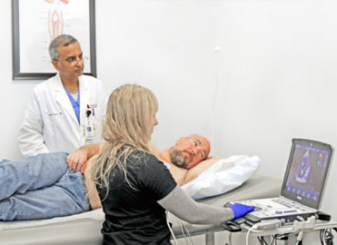
Echocardiogram
An echocardiogram uses high frequency sound waves to produce pictures of the heart. The ultrasound technician guides a small hand-held device called a transducer over the chest, that emits soundwaves. These sound waves bounce off internal substances and structures (in this case the heart), creating echoes that are picked up by the transducer. These are converted into moving images of the heart’s chambers, valves, blood vessels and pumping action. Combined with doppler technology, the speed and direction of the heart’s blood flow can also be measured at the same time.
Echocardiograms can help detect many aspects of heart disease including tumors, abnormal holes between the heart chambers (ASD & PFO), blood clots, and issues with the heart’s blood vessels, valves and pericardium (the heart’s outer lining).


